Macular Degeneration (AMD)
Macular degeneration is a condition that damages the macula, the central part of the retina. The macula is responsible for central vision and the ability to see detail.
When the macula is damaged, the eye loses its ability to see detail, such as small print, facial features, or small objects. The damaged parts of the macula often cause scotomas, or localized areas of vision loss. When you look at things with the damaged area, objects may seem to fade or disappear. Straight lines or edges may appear wavy.
The image on the right is what vision might look like with Macular Degeneration
What Are the Different Types of Macular Degeneration?
There are two types of the disease: dry macular degeneration and wet macular degeneration
Dry Macular Degeneration
Dry macular degeneration is a chronic eye disease that causes vision loss in the center of your field of vision. It is marked by the deterioration of the macula, which is in the center of the retina—the layer of tissue on the inside back wall of your eyeball.
Dry macular degeneration isn't associated with swelling and is the more common form of the disease. It doesn't cause total blindness but worsens your quality of life by blurring or causing a blind spot in your central vision. Clear central vision is necessary for reading, driving, and recognizing faces.
Symptoms:
The need for increasingly bright light when reading or doing close work
Increasing difficulty adapting to low light levels, such as when entering a dimly lit restaurant
Increasing blurriness of printed words
A decrease in the intensity or brightness of colors
Difficulty recognizing faces
A gradual increase in the haziness of your overall vision
A blurred or blind spot in the center of your field of vision
Hallucinations of geometric shapes or people in advanced stages
Dry macular degeneration may affect one or both eyes. If only one eye is affected, you may not notice much change in your vision because your good eye compensates for the weak one.
2. Wet Macular Degeneration
Wet macular degeneration is marked by swelling caused by leaking blood vessels that affect the macula. It almost always begins as dry macular degeneration, but it’s not clear what causes it to progress to the wet form.
Early detection and treatment of wet macular degeneration can reduce vision loss and, in some cases, improve vision. This is key to maintaining your vision with AMD.
Types of Wet Macular Degeneration:
Choroidal Neovascularization: Abnormal blood vessels grow from the choroid under and into the macula. These vessels leak fluid or blood, causing blurring and distortion of central vision.
Retinal Pigment Epithelial Detachment: Fluid leaks from the choroid and collects between the choroid and the retinal pigment epithelium (RPE), creating a blister or bump under the macula.
Symptoms:
Visual distortions, such as straight lines appearing wavy or objects appearing smaller or farther away
Decreased central vision
Decreased intensity or brightness of colors
Well-defined blurry spot or blind spot in your field of vision
Rapid worsening of symptoms
Hallucinations of geometric shapes, animals, or people in advanced cases
Diagnosis
Regular eye exams are the key to early detection of macular degeneration since symptoms may not be noticeable in the early stages. Early drusen (yellow deposits) can be seen during an eye exam before symptoms develop.
Risk Factors
Risk Factors You Can't Control:
Age
Race (Caucasians are at greater risk)
Genetics
Light eye color
Risk Factors You Can Control:
Smoking
High blood pressure
High cholesterol
Poor nutrition
Unprotected exposure to sunlight
Obesity
Sedentary lifestyle
An unhealthy lifestyle, such as smoking, poor nutrition, or limited exercise, could contribute to your risk of developing macular degeneration. Many risk factors are within your control to reduce your chance of getting the disease and to promote better health.
What You Can Do to Reduce Risk
You can lessen the risk of developing macular degeneration by reducing risk factors within your control, such as smoking and high blood pressure. No matter your age, you can incorporate the following guidelines into your life:
Quit smoking
Control high blood pressure
Control cholesterol levels
Improve nutrition
Wear 100% UV protective sunglasses
Maintain an ideal body weight
Exercise regularly
By following these guidelines, you may reduce your risk of developing macular degeneration and may be able to stabilize or slow the effects of vision loss if you have already been diagnosed.
Early Detection
People under 50 should have an eye exam every three to five years. Those with a family history of eye conditions or with medical conditions associated with eye disease, like diabetes, should have their eyes tested yearly, especially if over 65. If you notice changes in vision—such as blurriness—visit your eye doctor immediately.
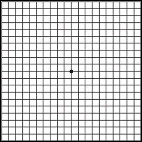
Normal Amsler Grid
Distortion of the normal pattern caused by wet macular degeneration
Treatments
Once dry AMD reaches the advanced stage, no treatment can prevent vision loss. However, treatment can delay and possibly prevent intermediate AMD from progressing to the advanced stage, where vision loss occurs.
The National Eye Institute's Age-Related Eye Disease Study (AREDS) found that taking a specific high-dose formulation of antioxidants and zinc significantly reduces the risk of advanced AMD and its associated vision loss.
AREDS Formulation:
500 mg Vitamin C
400 IU Vitamin E
15 mg Beta-carotene (or 25,000 IU Vitamin A)
80 mg Zinc (as zinc oxide)
2 mg Copper (as cupric oxide)
Who Should Take the AREDS Formulation?
People with intermediate AMD in one or both eyes
People with advanced AMD in one eye but not the other
The AREDS formulation is not a cure for AMD. It will not restore vision already lost from the disease but may delay the onset of advanced AMD.
Wet AMD Treatments:
Laser Surgery: Uses a high-energy light beam to destroy fragile, leaky blood vessels. It may also damage surrounding healthy tissue, and repeated treatments may be necessary.
Photodynamic Therapy: A drug called verteporfin is injected into the arm, traveling to the eye's abnormal blood vessels. A light shined into the eye activates the drug, destroying the vessels without harming healthy tissue.
Injectable Medications: Medications injected directly into the eye to treat abnormal blood vessels. These injections are usually spaced weeks or months apart and help slow vision loss.
Frequently Asked Questions
What is the dosage of the AREDS formulation?
500 mg Vitamin C, 400 IU Vitamin E, 15 mg Beta-carotene (or 25,000 IU Vitamin A), 80 mg Zinc, and 2 mg Copper.Who should take the AREDS formulation?
Those with intermediate AMD in one or both eyes or advanced AMD in one eye.Can people with early-stage AMD take the AREDS formulation?
No, it is not necessary for early-stage AMD. Regular eye exams can monitor progression.Can diet alone provide the same levels of antioxidants and zinc as the AREDS formulation?
No, the high levels are difficult to achieve through diet alone.Can a daily multivitamin provide the same benefits as the AREDS formulation?
No, multivitamins do not contain the high doses required. Consult your doctor before combining supplements.How is wet AMD treated?
Treatments include laser surgery, photodynamic therapy, and injectable medications to slow vision loss.
Macular degeneration is a leading cause of vision loss. Early detection, lifestyle changes, and appropriate treatments are key to managing the condition and preserving vision. Regular eye exams and proactive care are essential.
Click here to view a helpful document about Age Related Macular Degeneration.
How the medication is injected
Watch the videos below about how the eye works and Age Related Macular Degeneration and Dry Age-Related Macular Degeneration.


