Advanced Diagnostic Testing
At Carolina Macula & Retina, we utilize state-of-the-art diagnostic testing to assess and monitor retinal and macular conditions with precision. Advanced imaging allows us to detect eye diseases at their earliest stages, track disease progression, and determine the most effective course of treatment. Two of the most powerful diagnostic tools available in our office are Optical Coherence Tomography (OCT) and Fluorescein Angiography (FA). These non-invasive imaging techniques provide detailed insights into the structure and function of the retina, helping us deliver the highest level of specialized eye care.
Optical Coherence Tomography (OCT)
OCT is a cutting-edge, non-invasive imaging technique that provides high-resolution, cross-sectional images of the retina. Often referred to as “optical ultrasound,” OCT captures detailed images of sub-surface retinal structures, allowing for early disease detection and precise monitoring of retinal conditions.
Key Benefits of OCT:
Provides near-microscopic, real-time images of the retina.
Detects retinal abnormalities before they cause noticeable vision loss.
Requires no injections, contrast agents, or radiation, making it a safe and painless procedure.
How OCT Works:
OCT uses light waves to scan the retina, measuring the reflections from different retinal layers. The system filters out scattered light, isolating only the coherent reflected light to generate highly detailed, three-dimensional images. This allows for precise depth and structural analysis, helping to diagnose conditions such as macular degeneration, diabetic retinopathy, and glaucoma.
Fluorescein Angiography (FA)
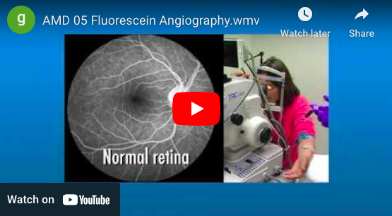
Fluorescein Angiography is a specialized imaging test used to evaluate the circulation of blood within the retina and choroid. This test helps detect conditions affecting the retinal blood vessels, such as macular degeneration, macular edema, diabetic retinopathy, and retinal vein occlusions.
How FA Works:
A small injection of a fluorescent diagnostic dye is administered into a vein in your arm.
As the dye circulates through the blood vessels in the retina, specialized photographs are taken.
These images help identify blockages, dye leakage, or poor circulation (ischemia) in the retina, providing essential information for diagnosis and treatment planning.
What to Expect After the Test:
Temporary yellowing of the skin for 24-48 hours.
Bright yellow or orange urine as the dye is eliminated.
Increased water intake can help flush the dye from your system faster.
Procedure Details & Safety:
The test typically takes about 30 minutes.
You may experience bright camera flashes during imaging.
Driving is not recommended after the test, so we suggest bringing a companion.
Mild side effects like nausea, headache, or dizziness are rare but possible.
Fluorescein Angiography is a safe and effective diagnostic tool that provides crucial insights into your retinal health. If you have any concerns before or after the test, our team is here to assist you.
By utilizing OCT and FA in our advanced diagnostic testing, Carolina Macula & Retina ensures that every patient receives the highest level of precision care. If you have been referred for diagnostic imaging or have questions about these tests, contact our office—we’re here to help protect and preserve your vision.
Fluorescein Angiography Video
Fluorescein Angiography Image


